Microscope What Is It Used For?
microscopes and microscopy
microscopes are used to magnify and observe tiny objects, organisms, and structures that are too small to be seen with the naked eye, allowing scientists and students to explore and understand the microscopic world.
a microscope is an instrument that can be used to observe small objects, even cells. the image of an object is magnified through at least one lens in the microscope. this lens bends light toward the eye and makes an object appear larger than it actually is.
microscopy is all about using tools to see things too small for the naked eye. at its core, microscopy involves magnifying tiny objects so we can study their details. this process has been essential in science for centuries.
microscopes work by using lenses to magnify small objects, making them visible and detailed. this basic concept has led to a wide range of microscopes, each designed for specific types of observation and research.
light microscopes are essential scientific instruments that allow us to see objects too small for the naked eye. these powerful tools use a series of lenses to magnify tiny specimens, revealing intricate details of cells, microorganisms, and tiny structures that would otherwise remain invisible.

light microscope observation
- in a typical light microscope, light passes through or reflects off a specimen, travels through objective lenseseyepiece lenses that further enlarge the image.
- this enables students and scientists to observe specimens at magnifications typically ranging from 40x to 1000x, opening up an entirely new world of observation.
- light microscopes are versatile tools used to examine a wide variety of specimens.
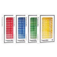
- students commonly use them to view prepared slides of plant and animal tissues, revealing cellular structures like cell walls, nuclei, and chloroplasts.
- they're perfect for observing microorganisms in pond water samples, including algae, amoebas, and paramecia.
- blood smears can be examined to see different types of blood cells.
- other common specimens include insect parts, textile fibers, thin sections of rocks and minerals, and even everyday items like salt crystals or human hair.
- the light microscope is ideal for viewing any specimen that is thin enough for light to pass through or that can be illuminated from above.

microscope use in biomedical research
microscopes are essential tools in biomedical research. these instruments help scientists, lab professionals, and researchers examine cells, tissues, bacteria, and other microscopic structures that are not visible to the naked eye.
the ability to see fine details plays a major role in understanding diseases, developing treatments, and carrying out accurate diagnoses.
different research applications require different types of microscopes. each type has specific features that support a particular function, such as magnification level, contrast techniques, lighting methods, or imaging capability.
- microscopy is an essential tool in many scientific disciplines.
- microscopes allow scientists to observe bacteria, viruses, fungi, and other microorganisms in great detail.
- this is crucial for understanding their behavior, structure, and how they cause diseases.
- observing living cells in real-time helps researchers study cellular processes, such as cell division and metabolic activities.
- these observations can lead to the development of new treatments and therapies for various diseases.
- additionally, they provide insights into how cells respond to different stimuli, which is vital for drug testing and development.
compound microscopes
- compound microscopes are the most commonly used microscopes in laboratories.
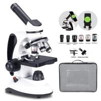
- they use two sets of lenses: objective and eyepiece, to magnify small samples.
- these microscopes are ideal for observing thin slices of tissue, blood smears, and bacteria.
- the compound microscope is a staple in many labs.
- it uses two sets of lenses—an objective lens and an ocular (eyepiece) lens—to magnify specimens.
- this type of microscope is excellent for viewing small details in thin samples like tissue slices or single-celled organisms.
- the compound microscope is typically used for observing objects at the cellular level.
applications:
- clinical sample analysis
- cell structure studies
- routine biological observations

fluorescence microscopes
- fluorescence microscopes are used to detect specific components within a sample by using fluorescent dyes.
- when exposed to certain wavelengths of light, these dyes emit fluorescence that can be observed and measured.
- this technique is highly valuable in molecular biology, immunology, and cell imaging.
- fluorescence microscopy: this method uses fluorescent dyes or proteins to label specific parts of the specimen.
- when illuminated with specific wavelengths of light, these labels emit light, creating high-contrast images of the targeted structures.
applications:
- dna and rna analysis
- cancer cell detection
- protein localization
phase contrast microscopes
- phase contrast microscopes are designed to observe transparent samples without staining.
- they enhance contrast by manipulating light waves, making it easier to view live cells and internal structures in their natural state.
- this type of microscope enhances the contrast of transparent specimens without staining.
- it’s especially useful for observing live cells and microorganisms in their natural state.
- phase contrast microscopy: this technique enhances contrast in transparent specimens without the need for staining.
- it works by amplifying differences in the refractive index of the sample, making it perfect for observing live cells and microorganisms.
applications:
- live cell monitoring
- bacterial motility studies
- cell culture inspection
stereo microscopes and dissecting microscopes
- stereo microscopes are used to observe the surface and structure of larger or opaque samples.
- they provide three-dimensional views at lower magnification.
- unlike compound microscopes, stereomicroscopes allow users to see the sample in depth, which is important when examining textures or performing dissections.
- also known as a dissecting microscope, the stereo microscope provides a 3d view of the sample.
- it’s perfect for observing larger, opaque specimens at lower magnifications, making it a favorite for dissection and detailed surface work.
- the dissecting microscope provides a lower magnification than the compound microscope, but produces a three-dimensional image.
- this makes the dissecting microscope good for viewing objects that are larger than a few cells but too small to see in detail with the human eye.
applications
- tissue dissection
- specimen sorting
- industrial quality control
electron and confocal microscopes
- electron microscopes:
- these microscopes use beams of electrons instead of light to achieve much higher magnifications and resolutions.
- transmission electron microscope (tem):
- tems pass electrons through thin samples to reveal internal structures in great detail.
- scanning electron microscope (sem):
- sems scan a focused electron beam across a sample's surface to create detailed 3d images of its surface.
- confocal microscope:
- confocal microscopes use lasers to create sharp, high-contrast images by focusing on a specific plane within the sample.
- confocal microscope:
- they are widely used in cell biology and materials science for their ability to produce clear, detailed images of thick specimens.
objects and organisms examined
- microscopy is used to examine tiny objects and organisms that are invisible to the naked eye.
- this includes anything from insect parts to plant cells, enabling detailed studies in fields like botany, zoology, and materials science.
- through these detailed examinations, scientists can better understand the structure-function relationship in various biological and material systems.
- this knowledge can then be applied to fields like agriculture, environmental science, and material engineering to develop innovative solutions.
- in cell biology, microscopes are indispensable for studying the complex architecture of cells.
- researchers use them to explore cell structures, such as the nucleus, mitochondria, and cytoskeleton, providing insights into cellular function and organization.
- detailed structural analysis helps in understanding diseases at the cellular level, such as cancer and neurodegenerative disorders.
- furthermore, it aids in the development of targeted therapies by revealing specific cellular pathways and mechanisms.
- microbiologists rely on microscopy to identify and characterize different microbes.
- this is vital for understanding microbial ecology, pathogenesis, and for developing new antimicrobial treatments.
- advanced techniques like fluorescence and electron microscopy offer detailed images of microbial structures and interactions.
- these detailed images can reveal how microbes interact with their environment and hosts, providing crucial information for controlling infections.
- additionally, they can uncover the mechanisms of resistance, helping to combat antibiotic-resistant strains.
magnification and microscope parts
magnification is at the heart of microscopy, allowing us to see tiny details that are invisible to the naked eye.
- objective lens & ocular lens:
- the objective lens is the primary lens that magnifies the specimen.
- microscopes often have multiple objective lenses with varying magnification levels.
- the ocular lens, or eyepiece, further magnifies the image produced by the objective lens, providing the final enlarged view that the user sees.
- eyepiece & nosepiece:
- the eyepiece is where you look into the microscope.
- it usually contains a lens that magnifies the image formed by the objective lens.
- the nosepiece is the rotating part that holds the objective lenses.
- by turning the nosepiece, you can switch between different objective lenses to change the magnification.
- condenser & light source:
- the condenser focuses light onto the specimen, improving illumination and contrast.
- the light source, typically a built-in bulb or led, provides the necessary light to view the specimen clearly.
- proper lighting is essential for obtaining high-quality images.
- mechanical stage & microscope slide:
- the mechanical stage is a platform that holds the microscope slide in place.
- it often includes knobs that allow precise movement of the slide to observe different areas of the specimen.
- the microscope slide is a thin piece of glass where the sample is placed for observation.
- focus knob:
- microscopes have two types of focus knobs: fine adjustment and coarse adjustment.
- the coarse adjustment knob moves the stage up and down to bring the specimen into general focus.
- the fine adjustment knob allows for precise focusing, ensuring that the image is sharp and clear.
magnification levels
- magnification refers to how much larger a microscope can make an object appear.
- the objective lens and the ocular lens work together to achieve this.
- the total magnification is calculated by multiplying the magnification power of the objective lens by that of the ocular lens.
- for example, if the objective lens is 40x and the ocular lens is 10x, the total magnification is 400x.
- microscopes can operate at different magnification levels.
- low power magnification (e.g., 4x or 10x) provides a broader view of the specimen, making it easier to locate areas of interest.
- high power magnification (e.g., 40x or 100x) zooms in on finer details, essential for studying intricate structures within cells and tissues.
- as magnification increases, the field of view (the visible area of the specimen) decreases.
- this means you see a smaller portion of the specimen but in greater detail.
- higher magnification also requires more light to maintain a clear image, so adjusting the light source and condenser is crucial when switching between magnification levels.
sample preparation and microscope selection
- preparing samples for microscopy involves various techniques to preserve and highlight the structures of interest.
- this might include staining, fixing, and sectioning the specimens.
- proper sample preparation is crucial for obtaining clear and informative images, whether it's for routine lab work or high-stakes research.
- effective sample preparation techniques can also enhance the visibility of specific components, aiding in more precise and accurate observations.
- these techniques are constantly evolving, improving the quality and reliability of microscopic analyses.
when selecting a research microscope, consider the following factors:
- sample type: transparent, stained, or opaque
- magnification needs: high magnification for cellular studies or low magnification for 3d views
- contrast method: brightfield, phase contrast, or fluorescence
- documentation needs: ability to capture and save images
- budget: cost-effective models or high-end systems with advanced optics
choosing the correct microscope improves the accuracy of research findings and the overall efficiency of laboratory operations.